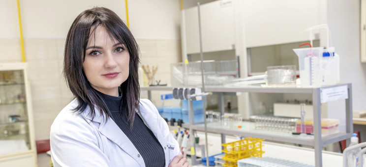
Joanna Nizol, DSc, Ph.D., Associate Prof. received a grant from the National Science Center as part of the SONATA BIS competition

Joanna Nizol, DSc, Ph.D., Associate Prof. from the Faculty of Chemistry of the Rzeszów University of Technology received a grant from the National Science Center as part of the SONATA BIS competition. The project aims to construct the first system for real 3D imaging of wide range of objects using mass spectrometry suitable also for the qualitative and quantitative analysis of biological samples at atmospheric pressure.
MSI technique
MSI is a molecular imaging technique that allows to visualize the spatial distribution of molecules. MSI can locate hundreds of chemical compounds, both endogenous (e.g., lipids, peptides, proteins) and exogenous (e.g., drugs, environmental pollutants) on the surface of an object in one experiment. It is an essential tool for analyzing the surface of objects of natural and synthetic origin. The currently used MSI methods allow locating elements in metals, polymers and semiconductors, as well as chemical compounds in biological samples. This family of methods have been used in biochemistry and medicine for many years, e.g., in research on the localization of metabolites secreted by microorganisms, tissue metabolites, drug pharmacokinetics or to search for disease biomarkers.
Currently, MSI is the best choice for structural-molecular analysis of biological samples. Traditional imaging methods, such as dye-staining or immunohistochemical staining in combination with fluorescence microscopy allow the visualization of tissue structures with high specificity and spatial resolution but are limited to a very small group of compounds. Other methods that identify chemical compounds, such as liquid chromatography coupled with mass spectrometry (LC-MS), require the extraction of analytes of interest from tissue homogenates, thereby losing important information about their spatial location in the tissue. MSI provides information on the concentration or amount of compounds and enables their localization on the surface of various solid samples. In a typical MSI experiment, the selected object is frozen, and then (for example) 10 µm sections are prepared using a cryotome and transferred to a plate compatible with the MS instrument, usually at room temperature. The plate with the sample is then placed in the mass spectrometer, where a series of measurements is made at various points on the surface under examination. The software creates an image from a dataset where a mass spectrum represents each pixel. The image can be generated for any selected m/z value from the pool of ions detected by the spectrometer with an abundance reflected in the displayed color.
MSI measurements are currently made in two dimensions (surface examination, 2D). Still, such information is not representative of the entire volume of the test sample, especially in the case of heterogeneous samples. Data from the whole sample volume can be crucial, especially in analyzing various biological samples where hidden (to optical examination) structures may be explored.
Molecular composition mapping in three dimensions is complicated, mainly for technical reasons. Obtaining three-dimensional representations of the distribution of a chemical compound may be accomplished by computer reconstruction from a series of 2D MSI data from many independently performed experiments for many thin sections of the object. In these measurements commercial instruments are used and the sample is in high vacuum conditions, which results in drying, curling, cracking of the sections, and loss of volatile metabolites. Moreover, living objects (e.g., human skin surface) cannot be tested.
The planned 3D MSI system will use ion source with three types of ionization. The optical system will consist of a pulse laser emitting far infrared and an innovative optical path. The proposed configuration will precisely control the ablation (removal) of selected layers of the object, which is crucial for 3D analysis. In the proposed 3D MSI system, samples will be analyzed without preliminary preparation, in a native or frozen state, and under atmospheric pressure. The technical solutions will enable the analysis of samples of relatively thick samples with a non-flat shape. By coupling this system with an ultra-high-resolution mass spectrometer, it will be possible to analyze the molecular composition. The test objects that will be analyzed as part of the grant are plant and animal tissues and tissues of human cancers. The results will be compared with those obtained by known methods.
A detailed description of the progress will be posted on the PolyTechnicLab team's website: https://www.facebook.com/profile.php?id=100090123766291 and https://twitter.com/PolyTechnicLab


