Researchers from WEiL develop a novel computer program for determining the quality of magnetic resonance images
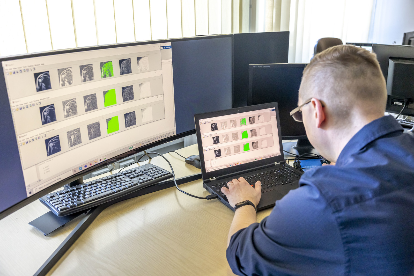
Scientists from the Department of Computer and Control Engineering at the Faculty of Electrical and Computer Engineering of Rzeszow University of Technology developed an automatic method for determining the quality of magnetic resonance images. The program can support radiologists or technicians by reducing the examination time.
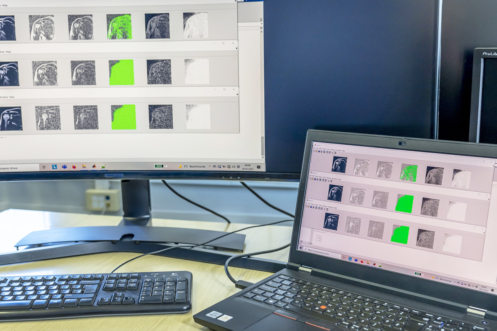
Magnetic resonance imaging (MRI) is a non-invasive examination that allows you to determine the condition of internal organs and tissues, as well as precisely capture pathological changes in the muscular, skeletal, and bone marrow systems. MRI is used, among others to diagnose diseases of the central nervous system, and the reliability of the results is of great importance. However, the poor quality of acquired medical images is often a problem that leads to the repetition of the examination.
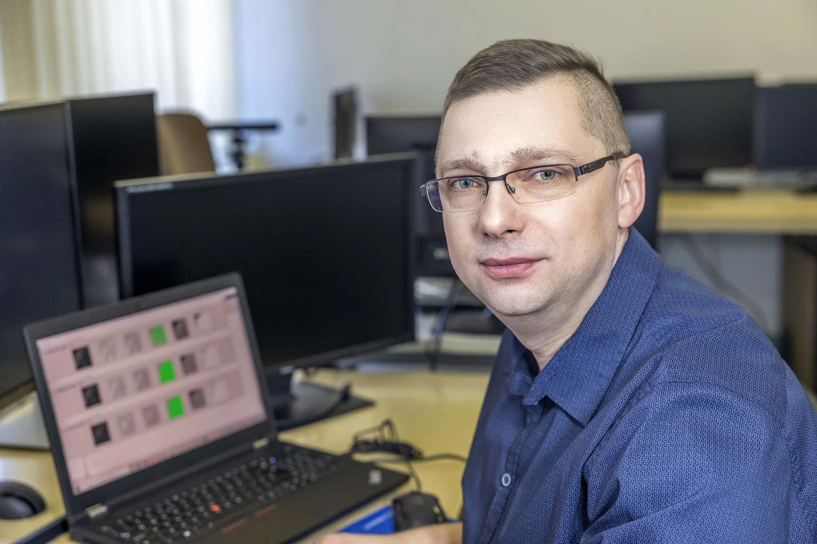
The idea for the project resulted from the observation of image quality problems that a radiologist or technician faces during an MRI examination. Mariusz Oszust, PhD, DSc, Eng., Associate Prof., from the Department of Computer and Control Engineering at the Faculty of Electrical and Computer Engineering of Rzeszow University of Technology emphasizes that the computer program developed at the university can support the technician during the procedure. The program allows for measuring the quality of acquired images in a way correlated with the average quality scores of a large group of radiologists. “Measuring the quality will allow for rejection of unsuitable scans, proposal of better scanner parameters, and possibly automatic scanner operation” – explains Mariusz Oszust, PhD, DSc, Eng., Associate Prof.
The quality of magnetic resonance images depends not only on the scanner parameters but also on the examined body location and the individual characteristics of the patient. The technician or radiologist conducting the examination is supported by the signal-to-noise ratio, and subjectively makes the decision on the eligibility of the images. The method developed by scientists from Rzeszow University of Technology for the assessment of the quality of magnetic resonance images is based on machine learning and may lead to automation and reduction of the examination time. The program offers additional information that the technician can use while considering the repetition of the he imaging sequence. This will automate the process and enable the selection of appropriate scanner parameters. Until now, solutions available on the market have not been offering similar functionality.
A computer program for determining the quality of magnetic resonance images can be used in institutions that perform this type of examination. This innovative project is also an interesting offer for manufacturers of imaging devices and phantoms for their calibration.
“The biggest challenge is to obtain a properly diversified database of MRI images and subjective assessment performed by a representative group of radiologists. Only thanks to sufficiently large data sets, the next stages of solution development, consisting in creating quality models with artificial intelligence methods, will be possible. The creation of such a database is both a financial and organizational challenge” – emphasizes Mariusz Oszust, PhD, DSc, Eng., Associate Prof.
The project was implemented thanks to funding from the Subcarpathian Center for Innovation (Podkarpackie Centrum Innowacyjności – PCI) in the 2nd call for grant proposals with amount granted of almost 90,000 PLN.
The work of Mariusz Oszust was financed by Subcarpathian Center for Innovation (Podkarpackie Centrum Innowacyjności – PCI), Teofila Lenartowicza 4 Street, 35-051 Rzeszów, Poland, under the grant no. N2_054, contract no. 32/PRZ/1/DG/PCI/2020 “Development of an automatic method for determining the quality of magnetic resonance images”; 2nd call for grant proposals.